邵阳学院附属第二医院 湖南 邵阳 422000 ; 2 、邵阳市中心医院 湖南 邵阳 422000 )
摘要:目的 以肺癌NCI-H1650细胞为研究对象,探讨二甲双胍(Met)与培美曲塞(Pem)联合对NCI-H1650细胞增殖抑制及机制。方法 MTT法测Met、Pem及联合干预对NCI-H1650细胞的影响;流式细胞术测Met、Pem及联合干预对NCI-H1650细胞周期的影响;PCR法测Met、Pem及联合干预后Bcl-2、Bax、Caspase-3的mRNA表达水平。结果 Met、Pem对NCI-H1650细胞增殖呈时间-浓度依赖抑制作用;联合干预增殖抑制作用要优于单药,联合干预作用表现为单纯相加。流式细胞术检测处理48小时的NCI-H1650细胞, Met干预后G期阻滞(P<0.01),Pem干预后S期阻滞(P<0.01),联合干预后G1和S期阻滞(P<0.01),Met及Pem均可诱导NCI-H1650细胞凋亡,且同一作用时间二者联合的凋亡率高于单药;与阴性对照组比,Met、Pem均抑制肺癌NCI-H1650 Bax表达,促进Bcl-2、Caspase-3表达,与阴性对照组和单药组比,联合组的协同作用更强,差异存在统计学意义(P<0.01)。结论 Met、Pem均能促进肺癌NCI-H1650细胞凋亡,抑制增殖,联合干预的抑制效果明显增强,作用机制可能是减少Bcl-2表达、增强Bax、Caspase-3表达。
关键词:二甲双胍;培美曲塞;肺癌NCI-H1650细胞
Inhibition of proliferation of NCI-H1650 cells by metformin combined with pemetrexed and its mechanism
Abstract: Objective To investigate the effect of metformin (Met) combined with pemetrexed (Pem) on the proliferation of NCI-H1650 cells. Methods MTT method was used to measure the effect of Met, Pem and combined intervention on NCI-H1650 cells; flow cytometry was used to measure the effect of Met, PEM and combined intervention on NCI-H1650 cell cycle; PCR method was used to measure the mRNA expression level of Bcl-2, Bax and caspase-3 after Met, Pem and combined intervention. Results Met and Pem had a time concentration dependent inhibitory effect on NCI-H1650 cell proliferation, and the inhibitory effect of combined intervention was better than that of single drug, and the combined intervention was simple addition. After 48 hours of treatment, NCI-H1650 cells were detected by flow cytometry. After treatment with Met and Pem, NCI-H1650 cells were blocked in G phase (P < 0.01), and in S phase (P < 0.01). Both Met and Pem could induce apoptosis of NCI-H1650 cells(P < 0.01). At the same time, the combined apoptosis rate was higher than that of single drug. Compared with the negative control group, met and PEM could inhibit NCI-H1650 cells Bax expression promoted Bcl-2 and Caspase-3 expression. Compared with negative control group and single drug group, the synergistic effect of combined group was stronger, and the difference was statistically significant (
P < 0.01). Conclusion Met and PEM can promote the apoptosis and inhibit the proliferation of NCI-H1650 cells. The inhibition effect of combined intervention is significantly enhanced. The mechanism may be to reduce the expression of Bcl-2, enhance the expression of Bax and caspase-3.
肺癌已经成为国内死亡率居于首位的恶性肿瘤,且呈逐年上升趋势[1]。因起病隐匿,发现时多已错失手术机会,全身化疗尤显重要[2]。二甲双胍(Met)是治疗糖尿病的基本药物,多项研究表明,其存在抗肿瘤效应,可抑制多种瘤细胞[3]。培美曲塞(Pem)是多靶点抗肿瘤药物,对肺癌疗效确切。本研究通过联合应用Met及Pem,探讨其对NCI-H1650的增殖影响及可能机制。为肺癌的个体化治疗提供理论依据。
1 材料与方法
1.1 材料 肺癌NCI-H1650细胞株购自上海细胞库,Met、Pem购自Solarbio公司,MTT、PI、BCA试剂盒、ECL试剂盒购自Sigma公司,PCR试剂盒购自Promega公司,Bax MAb、 Bcl-2 MAb、Caspase-3 MAb购自Santa Crus公司 。
1.2 方法
1.2.1 MTT法测NCI-H1650细胞增殖抑制率
对数期NCI-H1650细胞, 96孔板接种。培养后弃上清液, 分别用1、2、4mg/ml Met及1、2、4ug/ml Pem干预, 联合组分别用2mg/ml Met和2ug/ml Pem干预,设3复孔。阴性对照组, 添PBS , 24、48、72 h各加入 MTT液, 培养4 h, 以490 nm波长测光密度 (OD) 值,换算增殖抑制率 (%) 。
1.2.2 流式细胞仪测凋亡率及细胞周期的影响
对数期NCI-H1650细胞, 6孔板接种, 培养24 h。单一用药组向每孔细胞分别加入Met (2mg/ml) 、Pem(2ug/ml) , 联合用药组加入Met 2mg/ml及Pem 2ug/ml , 各2mL;阴性对照组不加药物, 培养48 h。标准处理。选用490 nm激发波长进行流式细胞仪检测, 重复3次。
1.2.3 RT-PCR检测Bax、Bcl-2和Caspase-3 m RNA的表达水平
对数期NCI-H1650细胞, 6孔板接种, 设双复孔, 培养24 h。分别加入Met (2mg/ml) 、Pem (2ug/ml)、Met (2mg/ml) 及Pem (2ug/ml) , 阴性对照组不加药, 培养48 h。抽取总RNA,反转录得cDNA。引物序列:Bcl-2基因, F-5'-CTTTTGCTGTGGGGTTTTGT-3, R-5'-GTCATTCTGGCCTCTCTTGC-3;Bax基因,F-5'-GGAGCTGCAGAGGATGATTG-3, R-5'-CCTCCCAGAAAAATGCCATA-3;Caspase-3基因, F-5′-AGGGGTCATTTATGGGACA-3′, R-5′-TACAC-GGGATCTGTTTCTTTG-3′;β-actin基因, F-5'-TGACGTGGACATCCGCAAAG-3,R-5'-CTGGAAGGTGGACAGCGAGG-3。按试剂盒说明操作RT-PCR, 反应条件:94℃变性5 min,扩增30个循环 (94℃60 s、57℃30 s、72℃60 s) , 72℃延伸5 min。重复3次, 分析灰度值。
1.3 统计学方法
用SPSS19.0统计数据,以均数±标准差( ) 表示,各组之间比较采用单因素方差分析,组间两两比较用t检验,P≤0.01为差异有统计学意义。
2 结果
Met抑制NCI-H1650细胞增殖, 同时间, 增加Met浓度, 抑制作用增强;同浓度, 延长Met作用时间, 抑制作用增强, 差异均有统计学意义 (P<0.01) , 见图1。
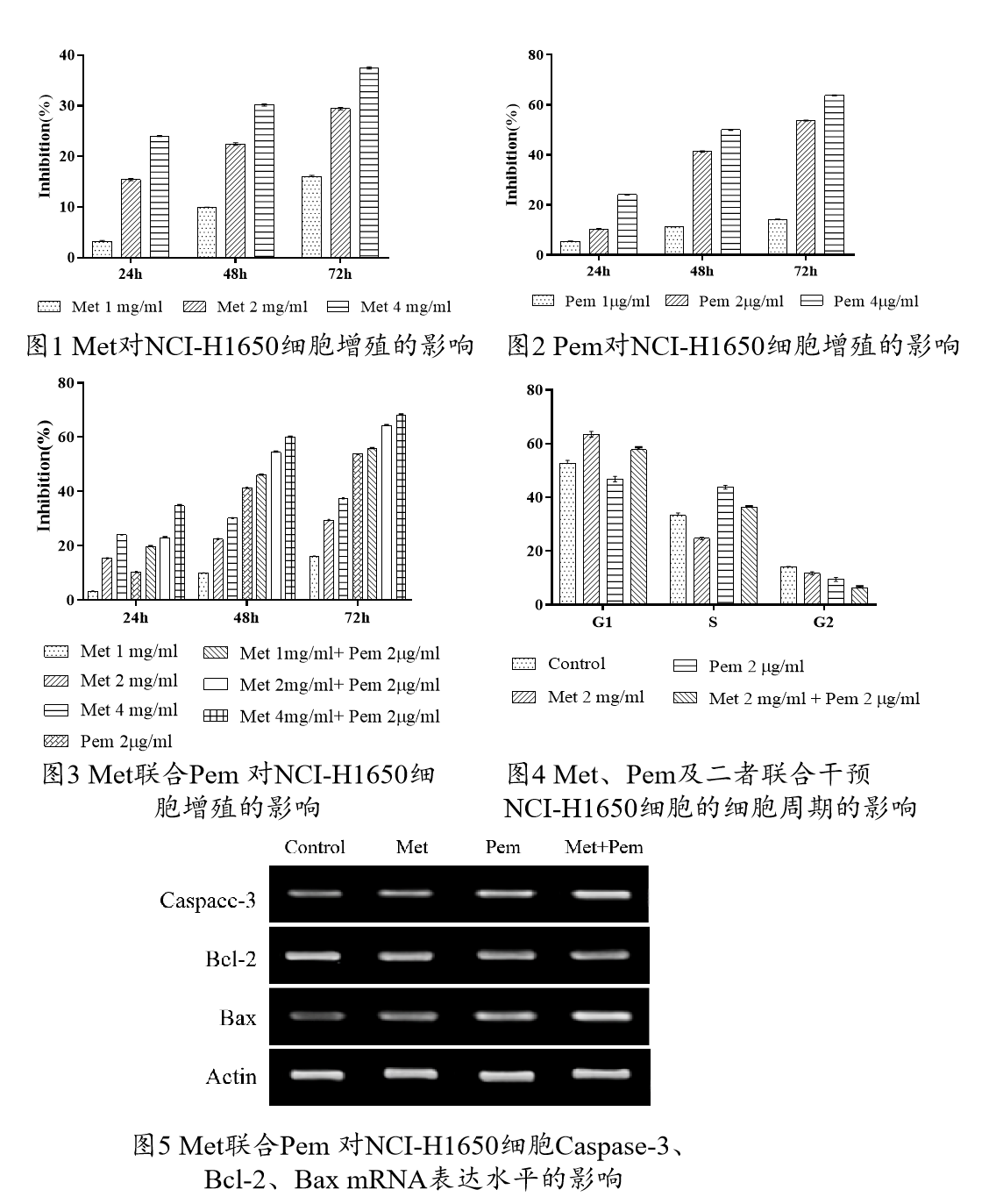
2.2 Pem对NCI-H1650细胞增殖的影响
Pem抑制NCI-H1650细胞的增殖, 同时间, 增加Pem浓度, 抑制作用增强;同浓度, 延长Pem作用时间,抑制作用增强, 差异均有统计学意义 (P<0.01) , 见图2。
2.3 Met联合Pem 对NCI-H1650细胞增殖的影响
Met与Pem联合对NCI-H1650细胞增殖有较强抑制作用, 延长作用时间, 抑制作用增强, 联合用药组抑制率与单一用药组比较, 差异均有统计学意义 (
P<0.01) , 见图3。
2.4 Met联合Pem 对NCI-H1650细胞周期和凋亡的影响
Met干预NCI-H1650细胞,G1期比例上升, 阻滞于G1期;Pem 干预,S期比例增加, 阻滞于S期;Met与Pem 联合可使G1、S期受到阻滞。差异有统计学意义(P<0.01), 见图4。
2.5 Met联合Pem 对NCI-H1650细胞Caspase-3、Bcl-2、Bax mRNA表达水平的影响
Met、Pem可增强NCI-H1650细胞Bax、Caspase-3 mRNA表达、抑制Bcl-2 mRNA表达, 差异有统计学意义 (P<0.01) 。Met与Pem联合作用效果更强, 与单药组比, 差异有统计学意义 (P<0.01) , 见图5。
3 讨论
肺癌是导致肿瘤患者死亡主要的原因,暂未找到治疗的特效方案。Pem对于肺癌的治疗存在确切疗效[4],但与紫杉醇等药物相比,没有明显优势,Met是糖尿病治疗的基本药物,研究发现其存在抗肿瘤作用,但机制不明[5]。
与单药物相比,联合用药可增强对NCI-H1650细胞的抑制作用,不同浓度的Met及Pem干预NCI-H1650细胞,增加作用浓度,不同浓度Met及Pem对细胞增殖的抑制作用均增强,联合用药后,表现出更强的抑制肿瘤细胞增殖能力,证实了Met联合Pem作用于NCI-H1650细胞具有相加作用。本实验发现,Met及Pem能使NCI-H1650细胞产生明显的凋亡诱导效应,而Met和Pem的联合使NCI-H1650细胞凋亡率最高,且本研究结果与上述研究者研究结果一致。可能经增强凋亡诱导效应来实现两药联合效应。Met阻滞瘤细胞于G1期,Pem阻滞瘤细胞于S期,共同诱导细胞凋亡。
Bcl-2及Caspase家族在细胞凋亡的调控过程中起重要作用[6]。Bax促进细胞凋亡,Bcl-2则抑制细胞凋亡,二者对立,Caspase-3起到中枢效应器作用[7]。三者相互影响。Bcl-2/Bax抑制Caspase-3表达,抑制细胞凋亡[8]。Caspase-3表达,抑制Bcl-2活力,促进细胞凋亡[9]。本研究采用RT-PCR检测肺癌NCI-H1650细胞Bcl-2、Bax、Caspase-3m RNA表达情况, 结果显示, 与阴性对照组比, 单药组Bcl-2 mRNA表达下调, Bax、Caspase-3 mRNA表达均上调, 联合组效果更明显, 提示Met、Pem可能通过下调肺癌NCI-H1650细胞Bcl-2mRNA表达水平, 上调Bax mRNA表达水平, 诱导细胞凋亡,同时, 上调Caspase-3 mRNA表达水平, 共同诱导凋亡。
综上所述, Met、Pem可能通过下调肺癌NCI-H1650细胞Bcl-2 mRNA表达, 上调Bax、Caspase-3 mRNA表达, 从而达到共同诱导凋亡的作用。
参考文献:
[1]Cukic V,Ustamujic A.Lung Cancer and Pulmonary Thromboembolism.Mater Socimed 2015;27(5):351-63.
[2]Willers H,Azzoli CG,Santivasi WL,et al.Basic mechanisms of therapeutic resistance to radiation and chemotherapy in lung cancer.Cancer J 2013;19(3):200-7.
[3]Massimo Moro,Elisa Caiola,Monica Ganzinelli,et al. Metformin enhances cisplatin-induced apoptosis and prevents resistance to cisplatin in co-mutated KRAS/LKB1 Non-Small Cell Lung Cancer (NSCLC)[J]. Journal of Thoracic Oncology,2018.
[4]Choi Mihong,Keam Bhumsuk,Ock Chan-Young,et al. Pemetrexed in the Treatment of Leptomeningeal Metastasis in Patients With EGFR-mutant Lung Cancer.[J]. Clinical lung cancer,2019,20(4).
[5]Salani Barbara,Maffioli Sara,Hamoudane Meriem,et al. Caveolin-1 is essential for metformin inhibitory effect on IGF1 action in non-small-cell lung cancer cells.[J]. FASEB journal : official publication of the Federation of American Societies for Experimental Biology,2012,26(2).
[6]肖丽娜,周先荣,张青华.Caspase-3蛋白在非小细胞肺癌中的表达及其与肿瘤细胞凋亡的关系[J].检验医学与临床,2019,16(14):2034-2036+2041.
[7]Li Xiaoli,Liang Liming,Yu Dee,et al. Gypenosides induces apoptosis in human non-small-cell lung cancer A549 cells via increasing the Bax/Bcl-2 ratio, caspase-3 and suppressing the NF-κB.[J]. Panminerva medica,2019.
[8]Thymoquinone induces apoptosis via targeting the Bax/BAD and Bcl-2 pathway in breast cancer cells. 2019, 46(3):411-417.
[9]Aneta Wnęk,Ewa Andrzejewska,et al. Molecular and immunohistochemical expression of apoptotic proteins Bax, Bcl-2 and Caspase 3 in infantile hemangioma tissues as an effect of propranolol treatment[J]. Immunology Letters,2017.
*基金项目:2018年邵阳市科技计划项目,项目名称:二甲双胍联合培美曲塞对肺癌NCI-H1650细胞的增殖抑制及可能机制(2018ZD02)

客服QQ:30444492琼网文【2021】1550-113号
增值电信业务经营许可证:琼B2-20210322
出版物经营许可证:新出发龙华出字第(2021)009号
广播电视节目制作经营许可证:(琼)字第00779号
版权所有 ©2002-2024 期刊网(www.qikanchina.com) 琼ICP备2021005105号